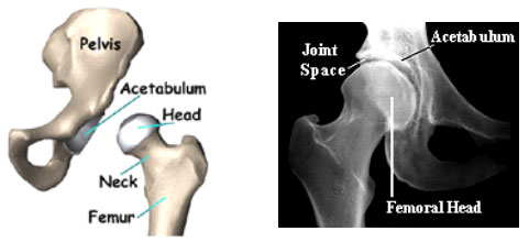
The hip joint is a ball-and-socket type of a joint formed where the thigh meets the pelvis. It consists of a socket called the acetabulum (part of the pelvis) and a ball called the head of femur (which is the top end of the thigh bone). The socket and the head of the femur are covered with covered with a smooth glistening white tissue called the articular cartilage which provides a low friction surface for movement. The joint is encased in a fibrous sheath called the capsule. The capsule has an inner smooth lining called the synovium which produces a lubricating fluid called the synovial fluid. The smooth cartilage along with the synovial fluid allows for frictionless movement at the joint interface. At the edges of the acetabulum is attached a fibro-cartilaginous rim called the acetabular labrum which helps to deepen the socket and adds to the stability of the joint. The joint is further stabilized by ligaments which are present both inside and outside the joint capsule. The hip joint is surrounding by a bulk of muscles which help to move the thigh at the hip joint.