What is articular cartilage?
Articular cartilage is the tough fibro-elastic tissue which covers the ends of long bones. It is a smooth, glistening white tissue which serves several functions – joint lubrication, stress distribution to underlying bone, and providing a smooth low-friction surface to the joint.
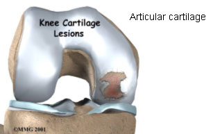

The articular cartilage is unique in that it lacks vascular, nervous and lymphatic elements which are present in most body tissues. It has a relatively low turnover rate and limited capacity to heal.
How do cartilage injuries occur?
Articular cartilage injuries may occur as a result of:
1. Acute impact (like a twisting injury or a road traffic accident)
2. Repetitive injury (like in sporting injury)
3. Secondary to a deformity in the knee, ligamentous instability in the knee or patello-femoral mal-alignment
4. Osteo-chondritis dissecans (OCD) – this is a condition which leads to detachment of an osteo-chondral fragment (articular cartilage with underlying bone). The exact cause of this condition is not well understood. This presents in the same way as any other cartilage injury.
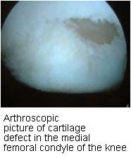
Though cartilage injuries can occur in any joint, it most commonly occurs in the knee joint. It tends to occur in the weight-bearing areas of the knee (commonest being the medial femoral condyle).
How does one diagnose cartilage injury?
Diagnosing cartilage injuries can be very difficult. The diagnosis is made on the basis of patient’s history of symptoms, physical examination, investigations like X-rays and MRI scans, and on Arthroscopy.
Patients may complain of pain on activity, swelling, catching, locking or giving way in their knee. There is no specific clinical test which can diagnose a cartilage injury. Plain X-rays may be normal and it may be necessary to take additional views like the 45 degree knee flexion weight-bearing view. X-rays may show a loose body inside the joint cavity. MRI scans may demonstrate a cartilage injury but it usually tends to underestimate the degree of cartilage damage. Arthroscopy allows for direct visualization of the cartilage injury, as well as assessment of associated injuries or pathology (like ligament instability). In many instances the cartilage injury is first diagnosed on arthroscopic surgery.
Treatment of articular cartilage injuries
In general the treatment depends upon the age of the patient, patient’s activity level and demands, degree of cartilage damage and location of the cartilage damage. The treatment options are conservative (non-surgical) and surgical.
Non-surgical treatment includes:
1. Oral medications (NSAIDs, Glucosamine and Chondroitin sulfate)
2. Physiotherapy
3. Braces (Unloader braces)
4. Injections (Corticosteroids and Hyaluronic acid derivatives like Synvisc)
5. Weight reduction
6. Activity modification
Non-surgical treatment is usually indicated in sedentary, low demand and elderly patients with degenerative changes.
Surgical treatment
The surgical options can be categorized as:
1) Palliative methods
a. Arthroscopic debridement & lavage
b. Thermal chondroplasty
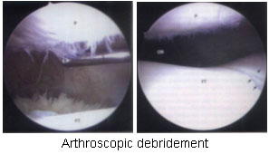
These methods only aim to remove the damaged cartilage and smoothen the joint surface, and as such will only give temporary benefit. These are indicated in low demand patients with small cartilage defects. These procedures are done entirely by arthroscopy.
2) Reparative methods
a. Microfracture
b. Subchondral drilling
c. Abrasion arthroplasty
These are known as marrow stimulation techniques. These methods involve surgical penetration of the subchondral bone (bone underlying the cartilage) which allows ingress of marrow cells which stimulates repair of the damaged cartilage with fibrocartilage. However the reparative tissue which forms at the cartilage injury site is not of the same quality as the damaged articular cartilage. These procedures are done entirely by arthroscopy.

3) Restorative methods
a. Osteochondral grafting (Mosaicplasty)
b. Autologous chondrocyte implantation (ACI)
These methods directly stimulate cartilage regeneration at the site of cartilage damage.
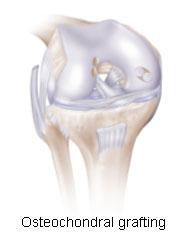
a. Osteochondral grafting
This involves implanting cylindrical plugs of bone and cartilage (osteochondral plugs) into the cartilage defect. These osteochondral plugs are taken from the non-weight-bearing parts of the femoral condyle. These plugs are then impacted into the area cartilage injury. The osteochondral plugs could either be taken from the patient himself (autograft), from a cadaver (allograft) or be artificial in nature (synthetic grafts). This procedure can be done entirely by arthroscopy.
b. Autologous chondrocyte implantation (ACI)
ACI is tissue engineering process which involves taking a small quantity of patient’s own articular cartilage from the knee and culturing these cartilage cells in a laboratory. These cultured cartilage cells (chondrocytes) are then implanted back into the cartilage defect during a second surgical procedure. This gives rise to healing of the cartilage defect with tissue which is bio-mechanically similar to the original cartilage. The first stage of this procedure (harvesting the cartilage from the knee) is done by arthroscopy. The second stage (implantation of the cultured chondrocytes) is performed a few weeks later and involves open surgery.

There are two different techniques available to implant the cultured cartilage cells. The conventional ACI technique (Carticel technique) involves the taking a flap of periosteum from the tibia (leg bone) and stitching it over the cartilage defect area, and injecting the cartilage cell culture underneath this periosteal flap. A modification of this technique (Chondron technique) involves mixing the cultured cartilage cells with fibrin gel which forms into an adhesive paste when applied on the cartilage defect. This avoids the need for an additional incision to remove the periosteal flap and obviates the need to suture the flap.


What’s new in the treatment of cartilage injuries?
The concept of harvesting stem cells and re-implanting them into one’s own body to regenerate organs and tissues has been tried in cartilage injuries as well. In particular, the mesenchymal stem cells have been shown in animal models to regenerate articular cartilage. These stem cells can be harvested from the patient’s bone marrow or fat cells. Recently there has been a published case report of successful cartilage growth in human knees using autologous mesenchymal stem cells. One will have to wait and watch for further developments in this direction.