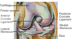
The knee joint is one of the largest and the most important joints in the body providing mobility and stability to the lower limbs. It is formed where the femur (thigh bone) meets the tibia (leg bone) and the patella (knee cap). The bones are encased in a fibrous sheath called the capsule. The ends of the femur and tibia and the undersurface of the patella are covered with a smooth glistening white tissue called the articular cartilage which provides a low friction surface for movement.
Interposed between the ends of femur and tibia are two crescent shaped fibro-cartilage structures each of which is called the meniscus. There are two such menisci in each knee – the medial meniscus (on the inner aspect of the knee) and the lateral meniscus (on the outer aspect of the knee). Their function is to act as shock absorbers for the joint and to increase the congruity of the joint.
The knee joint has four main ligaments (two inside the knee and two outside) which are made of tough fibrous tissue. The ligaments outside the joint are called collateral ligaments. There are two collateral ligaments – the medial collateral ligament (MCL) present on the inner aspect of the knee and the lateral collateral ligament (LCL) present on the outer aspect of the knee. The MCL prevents excessive outward bending and the LCL prevents excessive inward bending of the tibia on the femur. The ligaments inside the joint are called cruciate ligaments. There are two cruciate ligaments – the anterior cruciate ligament (ACL) which is present in the front of the knee and the posterior cruciate ligament (PCL) which is present at the back of the knee. The ACL prevents excessive forward translation of the tibia on the femur and the PCL prevents excessive backward translation of tibia on femur. The ligaments provide stability to the joint. Any injury to the knee thus causes some instability.
The knee joint is powered by two powerful groups of muscles – the quadriceps in the front of the thigh and the hamstrings on the backside of the thigh. The quadriceps is the all important muscle for knee function and is responsible for extending the leg on the thigh (straightening as well as lifting the leg up). The hamstrings are responsible for bending the leg on the thigh.